Thursday, December 20, 2012

Neuromuscular Junction

     Kendra and I made a poster about NMJ (Neuromuscular Junction) Here is a page about NMJ. We will also post a picture of our own poster: houseofmind:

The Neuromuscular Junction (NMJ) is a specialized synapse that serves to transmit electrical impulses (action potentials) from the motor neuron nerve terminal to the skeletal muscle. Basically, the NMJ allows for efficient and reliable communication between the motor neuron nerve and the muscles required for contraction and movement. The primary chemical messenger in this synapse, which consists of the presynaptic region (containing the nerve terminal), the synaptic cleft and the postsynaptic surface, is acetylcholine. These regions are defined by the differential localization of specific proteins, which underlie their distinct anatomical features and their physiological roles. 
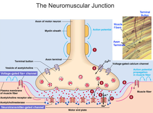
Now it’s time to briefly sum up what goes on in the NMJ, as shown in the diagram above. 
1. The action potential (or electrical impulse signal) reaches the nerve terminal in the presynaptic region. The hallmark feature of the nerve terminal is that it contains the synaptic vesicles, along with the proteins that help vesicle function. These vesicles are aligned near their release site, called an active zone. 
2. When action potentials reach the nerve terminal they activate calcium channels, which open up and facilitate the influx of calcium into the presynaptic terminal, which in turn commences the process of vesicular release into the synaptic cleft. 
3. The increase in intracellular calcium concentration triggers the fusion of the synaptic vesicles with the nerve terminal membrane. The mechanism of synaptic vesicle fusion involves conformational changes in multiple docking proteins both on the vesicle and the nerve terminal’s plasma membrane. 
4. Once fused with the nerve terminal membrane, the vesicle releases its contents into the extracellular space, also known as the synaptic cleft. The chemical or neurotransmitters (in this case, acetylcholine) released then bind to their corresponding receptors on the postsynaptic surface (also known as the motor end plate in the NMJ). 
5 & 6. Acetylcholine binds to its receptors and opens ligand-gated Na+/K+ channels. These structures are designed to optimize cholinergic neurotransmission in order to produce an end plate potential (EPP). The EPP is simply the net synaptic depolarization caused by the release of acetylcholine triggered by the nerve action potential. The EPP is a function of the miniature endplate potential (MEPP) amplitude, which represents the depolarization of the postsynaptic membrane produced by the contents of a single vesicle, and quantal content (number of transmitter vesicles released by a nerve terminal action potential. The EPP serves to open the voltage-gated Na+ channels in the postsynaptic region, which in turn results in an action potential that triggers muscle fiber contraction. These changes in the postsynaptic region potential result in muscle stimulation and contraction.
7. Acetylcholinesterase degrades acetylcholine so that it (choline) can be re-uptaked and recycled to produce new acetylcholine molecules. It’s activity terminates synaptic transmission. 
Sources:
Hughes, Benjamin W., et. al. 2006. Molecular architecture of the neuromuscular junction. Muscle & Nerve. 33(4): 445-461. DOI 10.1002/mus.20440
Motor Systems: Control of Movement and Behavior. 2008. Available at: http://www.colorado.edu/intphys/Class/IPHY3730/09motorsystems.html


NMJ is a specialized synapse. Its job is to transmit the electrical impulses throughout the body. It does so through the motor neuron nerve terminal. Once it leaves there, these impulses are sent all over the body. There are 7 steps for sending the electrical impulse, I will quickly describe all of them for you:
Step 1: There is an electrical impulse signal that has to reach the nerve terminal in the "presynaptic" region.
Step 2: When the impulses reach the nerve terminal, they activate calcium channels.
Step 3: The increase the calcium concentration will have to trigger the fusion of the synaptic vesicles with the nerve terminal membrane.
Step 4: Once this is fused with the nerve membrane, the vesicles will release its contents into the extra cellular space, this extra space is known as the "synaptic cleft."
Step 5&6: Acetylcholine binds to its receptors. It then will open ligand-gated channels. Next an end plate potential will be created.
Step 7: The final step, Acetylcholinesterase will degrade the acetylcholine so that the process can start over again and new acetylcholine molecules can be produced. 

Tuesday, December 18, 2012

Bone Structure

     If you like to study the human skeletal system, you will probably want to start with the support of the body. That would be the bones and bone structure. The best example I could start for beginners would be the femur. This bone is the longest and straightest single bone in the human body. It is also the strongest. If you break this bone, dont plan on walking again until it is completely healed or major problems could occur! the picture below is a human femur.
     Now, not all bones are solid in the human body. There are two specific types of bones when thinking of the structure. Cortical and Trabecular bones. Cortical bones are the hard and white bones you see that are the harder of the two. Trabecular bones are the honey comb looking holes inside the bone and are actually filled with marrow. Personally, I dont understand why the holes are considered "bone" but that is just me. It is important for bones to be strong to support our body weight and in some cases provide protection such as the skull and ribs. However, they must also be light enough to make movement possible. long bone consists of several sections:
  • Diaphysis: This is the long central shaft
  • Epiphysis: Forms the larger rounded ends of long bones
  • Metaphysis: Area between the diaphysis and epiphysis at both ends of the bone
  • Epiphyseal Plates: Plates of cartilage, also known as growth plates which allow the long bones to grow in length during childhood. Once we stop growing, between 18 and 25 years of age the cartilage plates stop producing cartilage cells and are gradually replaced by bone.
     Just to make sure you all understand this bone structure a little better, I will add a youtube video just for a little extra grasp of the concept! I will warn you now it is 8:42 minutes long but it is very informative and also explains everything about the picture above as well.